Updated Results of Singlet Oxygen Modeling Incorporating Local Vascular Diffusion for PDT
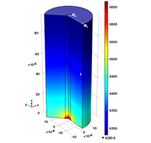
Introduction: Singlet oxygen (¹O₂) has a critical role in the cell-killing mechanism of photodynamic therapy (PDT). Therefore, in this study, the distance-dependent reacted ¹O₂ is numerically calculated using finite-element method (FEM). Herein, we use a model [Ref. 1] that has been previously developed to incorporate the diffusion equation for the light transport in tissue and the macroscopic kinetic equations for the ¹O₂ generation. In addition, the model includes the microscopic kinetic equations of oxygen diffusion from uniformly distributed blood vessels to the adjacent tissue. The blood vessel network is assumed to form uniformly distributed Krogh cylinders and the spacing (Rt) between vascular cylinders is varying between 20 and 60 μm. The cylindrical blood capillary has radius (Rc) in the range of 2-5 μm and a mean length (lz) of between 100 to 300 μm. Initial oxygen concentration [³O₂]₀ is considered to be 20 μM for tumor vasculature containing unstable endothelium and leaky vessels. This is achieved by setting the oxygen pressure (Pts) at aortal entrance of the blood vessel to be 50 mmHg, lower than what was used for normal tissue at 80 mmHg. The blood velocity in capillary (vz) is also considered to be in a broad range of 50-750 μm/s.
Use of COMSOL Multiphysics® software: The forward calculation for the singlet oxygen generation model incorporating the macroscopic kinetic equations was done using COMSOL. Within COMSOL, the finite-element calculation was implemented by varying the input parameters, such as light fluence rate.
Results: When the mean length of the capillary increased from 100 to 400 mm, the resulting blood perfusion rate (g) changes from 4.05 to 0.99 μM/s following roughly 1/lz for otherwise the same conditions of Rc= 2.5 and Rt = 60 μm. When the oxygen pressures (Pts) are increased from 10 mmHg to 100 mmHg, g changed from 0.13 to 0.89 μM/s for otherwise the same conditions of Rc = 2.5 and Rt = 60 μm. When the blood velocity increased from 50 μM/s to 750 μM/s, g increased from 1.24 to 7.55 μM/s for otherwise the same conditions of Rc = 2.5 and Rt = 60 μm. When the metabolism consumption of tissue increased from 0.9 to 16 μM/s, g decreased from 2.32 to 12.24 μM/s for otherwise the same conditions of Rc = 2.5 and Rt = 60 μm.
Conclusion: Several improvement of the model formulations have been made to prove the validity of the formulation of the oxygen perfusion term, g(1-[³O₂]/[³O₂]₀), used in the macroscopic singlet oxygen model. The variation of g is between 0.13 μM/s to 294 μM/s among the feasible parameters obtained from literature.

Download
- penjweini_paper.pdf - 0.8MB
- penjweini_abstract.pdf - 0.02MB
